Stage 01 of 05 · weeks 18-22
The 20-week scan: what heart findings actually mean
The mid-pregnancy anatomy scan is a general survey of the whole fetus. The heart gets a few specific views. Understanding what those views can and cannot show is the key to not catastrophizing - or dismissing - what you were told.
The short answer
At the 20-week scan the sonographer looks at four standard views of the heart. If something was flagged, that is a referral for a closer look - not a diagnosis. Most flagged findings turn out to be minor or resolve. A normal scan is a good sign but not a lifetime guarantee. Write down the exact words used; that word is what you will search tonight.
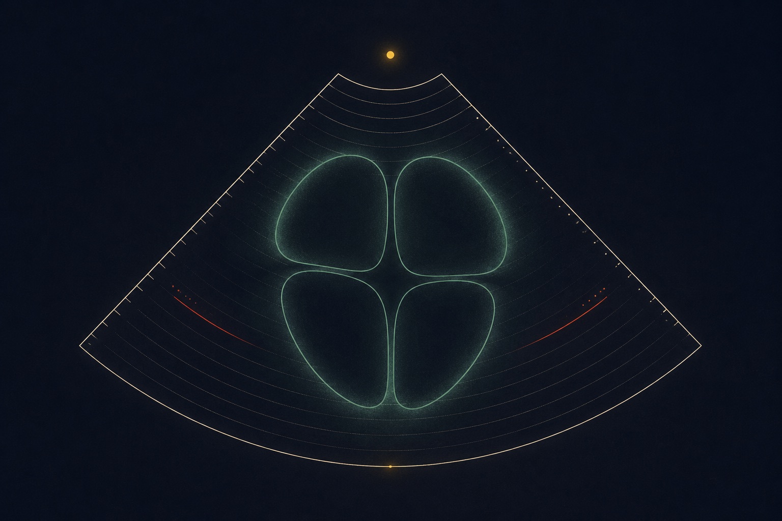
What actually happens at the 20-week scan?
A sonographer examines the fetus systematically. For the heart, international guidelines define a basic examination: the four-chamber view (all four chambers, the septum between them, and the crux), plus outflow-tract views (the left and right ventricular outflow tracts, and where the great arteries cross). Some countries add the three-vessel-trachea view as standard.
The scan typically takes 20-40 minutes in total. The heart portion is a matter of minutes. The sonographer is looking at anatomy - structure - not function.
Evidence
The standard basic cardiac screening examination is defined by the International Society of Ultrasound in Obstetrics and Gynecology: four-chamber view plus outflow tracts; detection rates for major CHD roughly double when outflow-tract views are added. Source: ISUOG Practice Guidelines (updated): fetal cardiac screening, UOG 2023.
They said they "saw something." Does that mean something is wrong?
The screening scan is deliberately sensitive: it is designed to flag anything that deserves a closer look, accepting that many flags will turn out to be nothing. Three honest facts:
- Most structures seen "suspiciously" at screening normalize or prove minor at the focused scan. Common examples are a bright spot in the ventricle (echogenic intracardiac focus) and small size differences that fall within normal range on repeat measurement.
- Some findings are genuinely important. Screening detects a majority - not all - of major structural heart disease. That is why referral to fetal echocardiography exists.
- A normal screening scan is a good sign, not a guarantee. Some heart problems develop or become visible later in pregnancy, which is why some units re-look at the heart in the third trimester.
Who will I be dealing with now?
A sonographer performs most screening scans. In many countries they are not permitted to discuss findings directly - they pass images to an obstetrician or radiologist. That silence is protocol, not concealment. The obstetrician or a midwife then explains what was seen and makes any referral.
What happens in the first 48 hours?
Research on parents' education needs after a CHD diagnosis found the period immediately after a finding is when information retention is lowest - and anxiety highest. Three things consistently help:
- Write down the exact words used, or ask for them in writing.
- Confirm when and where the fetal echocardiogram will happen, and whether it needs to be sooner.
- Arrange for one support person to attend that appointment with you.
Evidence
Parent-education recommendations - staged disclosure, question lists, support-person involvement - drawn from a crowdsourced study of families post-CHD diagnosis. Source: BMC Pregnancy & Childbirth 2023.
What to ask
Bring these to your next appointment
- Which exact view or structure was flagged?
- Is this finding known to sometimes resolve or stay minor?
- How soon should the fetal echocardiogram happen - days or weeks?
- Who will perform it, and are they a fetal-cardiology specialist?
- Does anything change in my routine care before then?
- Can I have the report in writing, with the words used?